
Complex models at NPSC
Complex cellular assay services for spheroids, primary cells, tissue models and iPSCs, with live imaging, multi-omics readouts and scalable screening
Biological assays are moving beyond standard laboratory cell models. The NPSC has experience in developing and screening complex cellular assays in well plate format. We can obtain multiple readouts from a single experiment, for example a live-imaging readout followed by protein or RNA extraction on the same samples. We have worked with:
- spheroids
- primary human samples
- physiologically relevant tissue models
- iPSCs and their derivatives (in collaboration with the HPSCF)
Get in touch using our enquiry form, or e-mail [email protected]
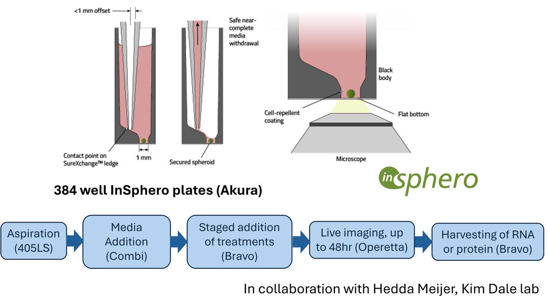
Live imaging and analysis of spheroids in 384 well plates
Oscillations of expression in response to Wnt signalling are observed over a 48hr period. The InSphero plates (Akura); together with our liquid handling capabilities, facilitate precise, staged media changes and washes of the spheroids. This is followed by extraction of RNA or protein for downstream applications once live imaging is finished.
Physiologically relevant lung cell model
Air-liquid interface (ALI) cultures, grown on transwells, generate a differentiated, physiologically relevant model of airway cells. The presence of Muc5AC, NGFR, and acetyl-Tubulin positive cells confirms the presence of Goblet, Ciliated, and Basal cells. The presence of E-cadherin and ZO-1 confirm the epithelial properties of the cells.
Potential assays with this model include:
- bacterial or viral infection assays
- co-culture assays (e.g. immune cell infiltration)
- cilia-beating assays
Potential readouts include microscopy, proteomics (150ug protein can be obtained from a single well of a 96 well plate), RNA seq, or apical and/or basal secretion.
Other tissues can be discussed and developed.
