High content analysis at NPSC
Phenotypic profiling and Cell Painting services deliver high-content imaging, MOA prediction and drug screening
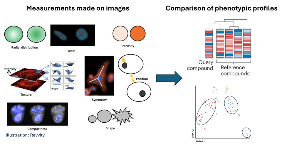
Phenotypic profiling, or high content analysis, aims to provide a holistic, whole cell phenotype to report on the function of a perturbation in a non-biased way. The approach required 1000s of measures to be made on every cell in an image. We use the Cell Painting assay, which uses 6 standard stains against sub-cellular components. Image measurements are compared with known (reference) treatments; similarity of these profiles are used to identify treatments inducing a similar effect.
We have experience running the Cell Painting assay in multiple cell types:
- standard cell lines (HeLa, U2OS, A549, HepG2 cells)
- iPSCs (in collaboration with HPSCF)
- human spermatozoa
- malaria parasites
We can use phenotypic profiles to predict compound MOA, screen for compounds which rescue an undefined cellular phenotype, or to generate the data to inform a machine learning model.
Get in touch using our enquiry form, or e-mail [email protected]
Overview of Cell Painting
The Cell Painting imaging panel includes: DAPI, to visualise DNA and nuclei; Concanavalin A for ER; Syto14 for nucleoli and cytoplasmic RNA; Phalloidin for Actin; Wheat Germ Agglutinin for Golgi and plasma membrane; MitoTracker for mitochondria.

Measurements are made on each of the fluorescence reporters to produce a phenotypic profile for each treatment. Similarity in profiles help to identify treatments with similar effects.
Applications of cell painting
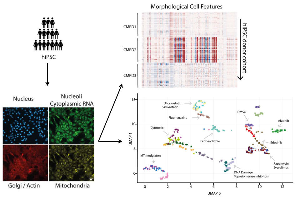
iPS Cell Painting uncovers differential drug responses
In collaboration with the HPSCF, Professor Jason Swedlow and Professor Angus Lamond, we established Cell Painting in undifferentiated iPSCs.
A panel of drugs were screened against a cohort of 28 iPS cell lines (HipSci), and responses measured by Cell Painting. Variable phenotypic responses between hiPSC lines were detected with a wide range of clinically approved drugs in use across multiple disease areas. Information on mechanisms of drug-cell interactions underlying the observed variable responses was derived using proteomic analysis.
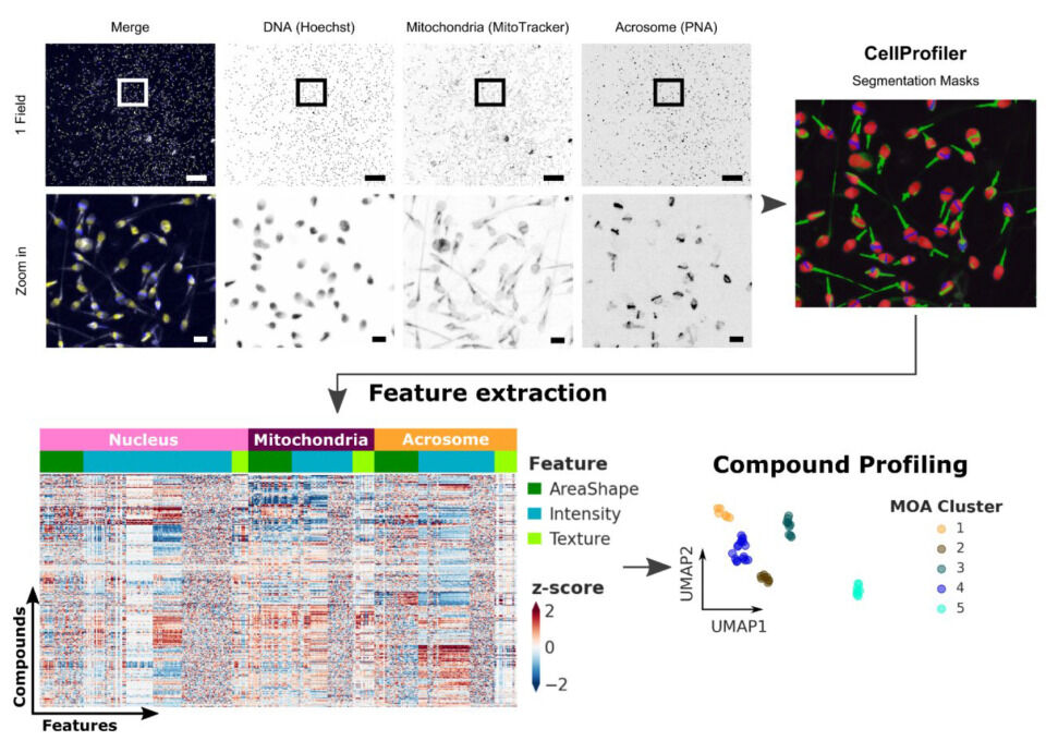
Sperm Cell Painting
With our colleagues in the Faculty of Health (Professor Chris Barratt) and the Drug Discovery Unit, we have modified the standard Cell Painting protocol to be suitable for sperm cells.
The different components important to sperm cell function are represented in the dye panel, which includes Hoechst to visualise DNA and the nucleus; MitoTracker to visualise the mitochondria; and PNA to visualise the acrosome, a sperm-specific structure required for fertilisation of an egg.
Object segmentation and feature extraction are performed using the same tools as for standard cell painting.
Read more about Sperm Cell Painting.
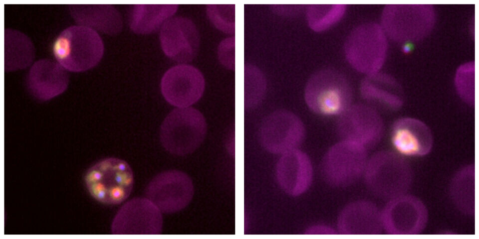
MOA prediction for Malaria treatments
With Professor Marcus Lee (BCDD), Medicines for Malaria Venture (MMV), and LPixel, we are developing a method to predict the MOA of Malaria treatment using Cell Painting.
We have adapted the Cell Painting assay to report on important aspects of Plasmodium cell function utilising a panel of organelle dyes.
Read the press release