
Location
Dundee Imaging Facility
We provide light and electron microscopy resources, with a central hub in Life Sciences and facilities in Science and Engineering and on the Ninewells campus.

Dundee Imaging Facility
School of Life Sciences
University of Dundee
Dundee
DD1 5EH
Accessibility
View accessibility information for the Medical Sciences Institute (Life Sciences) at AccessAble
View accessibility information for the Wellcome Trust building at AccessAble
View accessibility information for the School of Medicine (Ninewells Campus) at AccessAble
View accessibility information for the Harris Building at AccessAble
Please get in touch if you would like to discuss your project with us
About
The facility has a central hub in the School of Life Sciences, incorporating:
- light microscopy
- tissue imaging
- the Physics and Life Science (PaLS) lab
- image analysis
- sample preparation facilities
We have an additional Light Microscopy facility at the Ninewells campus, School of Medicine and our Analytical Electron Microscopy facility is housed in the School of Science and Engineering. All of our technology is accessible by all staff in the University of Dundee on an equal basis.
The facility is overseen by a cross-School Advisory Steering Group.
Histology
Our histology service supports users in all aspects of sample preparation and provides expert advice, training and practical support in a variety of histological techniques. As well as conventional paraffin wax histology we can also cut frozen thin and thick sections.
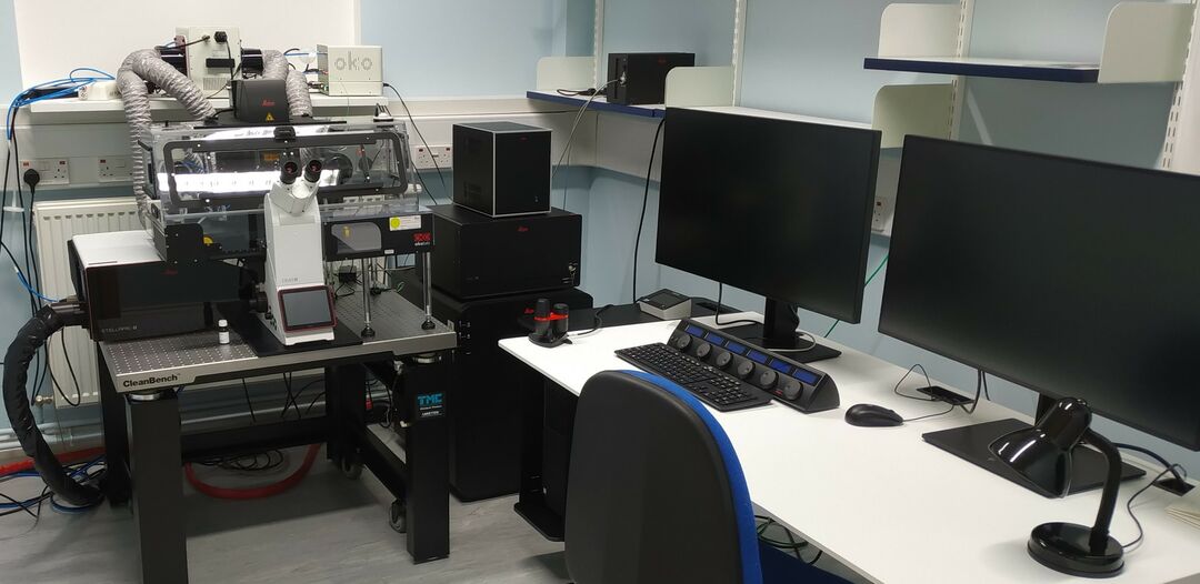
Light microscopy
The facility houses a range of advanced fluorescence imaging systems, all are equipped for live cell imaging. We cover a wide range of applications including multi-wavelength 3D imaging of fixed material, live imaging of cells, tissues and embryos, FRAP, FRET, DNA damage and laser ablation.
- Widefield Deconvolution Microscopy (DeltaVision, Nikon and Zeiss)
- Laser Scanning Confocal and Multiphoton microscopy (Zeiss and Leica)
- Laser Micro- dissection (Zeiss)
- Total Internal Reflected Fluorescence Microscopy – TIRF (Nikon)
- Spinning Disk (Zeiss and Nikon)
- Time Correlated Single Photon Counting Fluorescence Lifetime Imaging – FLIM (Zeiss and Leica)
Image analysis
Imaging experiments are quantitative assays, so expertise in computational data analysis is a critical component of the Dundee Imaging Facility’s capabilities. We have a suite of 7 high performance PCs running a range of commercial and open source software supported by an Image Analyst. Our Image Analyst supports several complementary software platforms (Fiji/ImageJ, OMERO, Imaris, Volocity, CellProfiler, Zen, LAS X, Nikon Elements, RStudio) and provides expertise in many different analysis frameworks (including Java, Python, Matlab and R). Custom tools can be developed where required.
Electron microscopy
Our electron microscopes are housed in the School of Science and Engineering (Harris Building) and comprise:
- SEM - JEOL 2200FS with EDX
- JEOL - 1200 TEM.
Support is available for complex techniques, including ultramicrotomy, immunogold labelling and x-ray micro analysis. A full range of sample preparation equipment is available for SEM and TEM
Supporting your research
If you are internal to the University, you can find out more details, including booking information and pricing, on our internal SharePoint site.
If you are an external client and you’d like to find out more about how we can support your research, please get in touch to discuss your project further.
All work with external clients is undertaken in accordance with our standard Terms & Conditions.
People
| Name | Role |
|---|---|
| Dr Graeme Ball | DIF Specialist |
| Ian Newton | Senior Technician |
| Kirsten McLeod | Senior Technician |
| Dr Paul Appleton | DIF Manager and Expert |
We provide a fast-paced, collegiate and supportive environment for our staff and students.