
Anatomy of the Inner Ear
Overview
Content Expert:
- Dr Patrick Spielmann
Content Reviewers:
- Dr Penny Lockwood
- Dr David Stewart
Medical Artist
- Annie Campbell
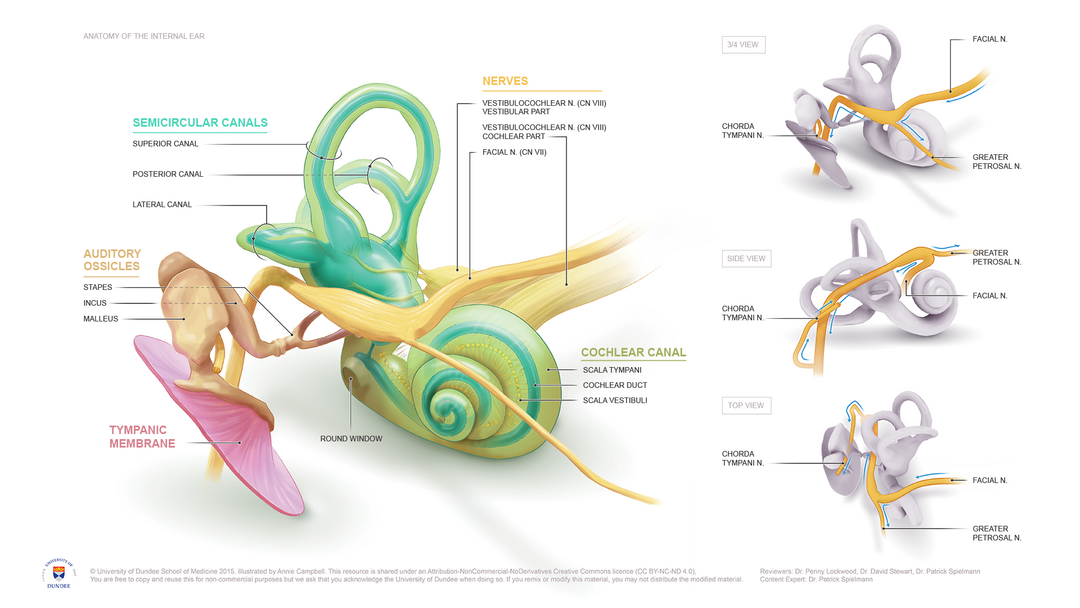
Certain aspects of this model were created from segmented MRI data*, making this a highly accurate representation of the tympanic membrane, facial nerve, ossicles and vestibular system.
This work “Anatomy of the Inner Ear”, is a derivative of “3D Ear” by W. Robert J. Funnell, PhD; Sam Daniel, MD, CM; and Daren Nicolson, MD, CM at McGill University, used under CC BY-NC-SA 1.0. “Anatomy of the Inner Ear” is licensed under CC BY-NC-SA 4.0.
*The vestibulocochlear nerve was not derived from MRI data, however heavily referenced
Final Product
You are free to copy, reuse and remix this for non-commercial purposes but we ask that you acknowledge the University of Dundee as well as publish any remixed work under the same share-alike license as the original authors.