
News
New Live-Imaging Tools created through multi-lab collaboration with Dundee's Pluripotent Stem Cell Facility
A new collaborative study, published in the journal Development, reports and validates new tools and techniques with which to interrogate human cell behaviour and cell biology using live cell imaging approaches.
Published on 9 December 2025


Publication authors (anti-clockwise from top): Alwyn Dady, Timothy Sanders, Greg Findlay, Kate Storey, Lindsay Davidson, Sophie Rappich, Nicolas Loyer, Jens Januschke
These new tools will be valuable for many researchers in the expanding fields of human reproduction and embryonic development as well as in disease modelling. It is the result of a multi-lab collaboration between the Storey, Findlay, Januschke groups in the School of Life Sciences, Timothy Sanders (University of Pittsburgh) and the Human Pluripotent Stem Cell Facility led by Dr Lindsay Davidson.
Professor Kate Storey explains, “We used a genetic engineering approach (mediated by PiggyBac transposase) to introduce fluorescent markers for the cell membrane, nucleus or actin cytoskeleton into human pluripotent stem cells or directly into in vitro generated human neural tissue – an approach made possible by the expertise and support of the Human Pluripotent Stem Cell Facility. This engineering approach is fast, and the reporters are advantageous for live cell imaging, visualising protein dynamics inside individual cells within tissues - including actin cytoskeleton dynamics in brain progenitors that form small neural rosettes”.
Co-author Nicolas Loyer’s microscope image from the study was selected as the front cover image for the November 2025 edition of Development. It shows a neural rosette expressing a fluorescent marker for the actin cytoskeleton.
Read the study in full here:
Dady et al. 2025. ‘Engineering fluorescent reporters in human pluripotent stem cells and strategies for live imaging human neurogenesis’ Development PMID: 40748212 DOI: 10.1242/dev.205082
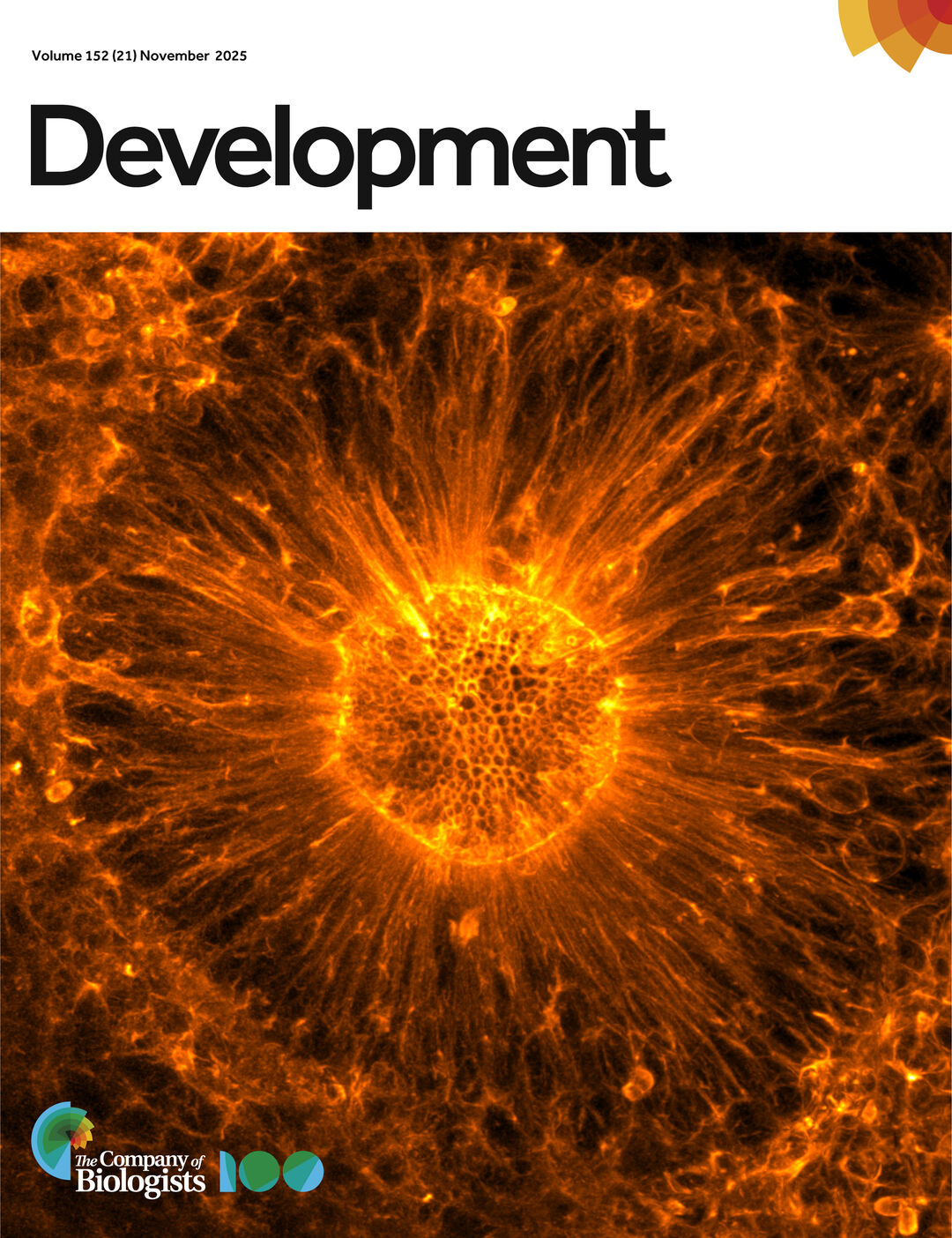
Nicolas Loyer’s microscope image of a neural rosette from the study, Dady et al. that was selected as the front cover image for the November 2025 edition of Development.